We’re excited to announce that Dr. Jeffrey M. Martinez is now offering Genicular Artery Embolization (GAE)—a groundbreaking, minimally invasive treatment for knee osteoarthritis pain.
At Espada Vascular, we specialize in the diagnosis and treatment of complex venous diseases. Our team, led by Dr. Jeffrey M. Martinez, is dedicated to providing advanced vascular care focused on prevention, preservation, and life-saving outcomes. We understand that conditions affecting your veins can significantly impact your daily life, causing discomfort, swelling, and more serious health complications. We offer personalized, minimally invasive treatment plans designed to restore healthy blood flow and improve your overall quality of life right here in San Antonio.

Venous diseases encompass a range of conditions that affect the veins in your body, primarily those in your legs and pelvis. Your veins are responsible for carrying oxygen-poor blood back to your heart. When these vessels become damaged, narrowed, or blocked, it can lead to blood pooling, increased pressure, and a variety of uncomfortable symptoms. It is crucial to address these issues promptly to prevent disease progression and maintain long-term wellness.
We provide comprehensive care for several specific venous conditions. Our goal is to educate our patients and offer the most effective treatments available.
Please note that while we treat a wide array of complex vascular issues, we do not provide services for spider veins.
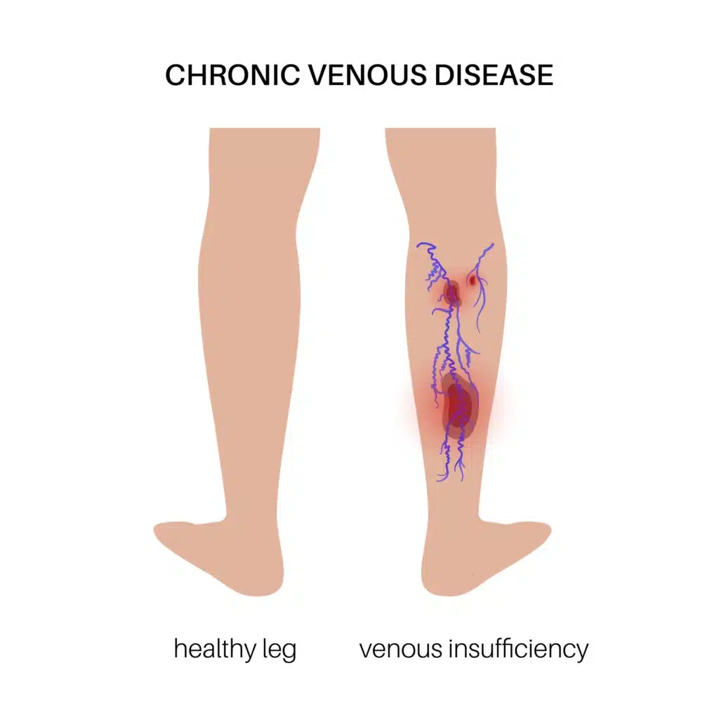
Venous insufficiency, also referred to as chronic venous insufficiency (CVI), is a condition in which the veins in the legs are unable to pump enough blood back up to the heart. This occurs when the one-way valves inside the veins become damaged or weakened, allowing blood to flow backward and pool in the lower extremities. Over time, this pooling increases pressure in the veins and can lead to a range of progressive symptoms and complications. According to StatPearls via the National Institutes of Health, chronic venous insufficiency is one of the most common vascular conditions, with a wide spectrum of clinical presentations.
The symptoms of venous insufficiency can range from mild to severe and often worsen over time if left untreated. Common signs include persistent leg swelling, a feeling of heaviness or fatigue in the legs, aching or cramping, skin discoloration or thickening around the ankles, and the development of varicose veins. In advanced cases, the skin may become leathery or develop a condition called lipodermatosclerosis, and open wounds known as venous ulcers may form.
Diagnosis typically involves a physical examination and duplex ultrasound imaging to assess blood flow and identify any valve dysfunction or reflux in the veins. Treatment is tailored to the severity of the condition. Mild cases may be managed with compression therapy, leg elevation, and lifestyle modifications. For more advanced venous insufficiency, minimally invasive procedures offer effective relief. Vein ablation, which uses radiofrequency or laser energy to close the diseased vein, is a widely used and well-established approach.
Varithena, an FDA-approved injectable foam, is another option that works by chemically closing incompetent veins, particularly those of the great saphenous system, without the need for surgery or thermal energy. A comprehensive review published in PubMed highlights that early intervention for chronic venous insufficiency significantly reduces the risk of disease progression and improves patient outcomes.
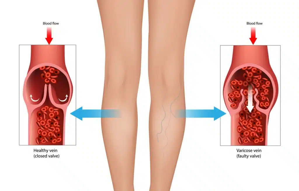
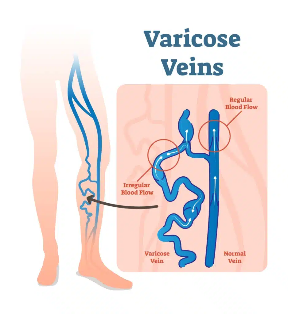
Varicose veins are enlarged, twisted veins that most commonly appear in the legs and feet. They occur when the small, one-way valves inside the veins become weak or damaged, allowing blood to flow backward and pool in the vein. This causes the vein to stretch and bulge, becoming visible just beneath the skin. Varicose veins are one of the most common early manifestations of venous insufficiency.
While often considered a cosmetic concern, varicose veins can also cause physical discomfort, including an aching or heavy feeling in the legs, burning, throbbing, muscle cramping, and swelling in the lower legs. Symptoms may worsen after prolonged periods of sitting or standing. It is important to note that while we treat symptomatic varicose veins associated with broader chronic venous disease, we do not provide services for spider veins, which are smaller, web-like veins closer to the skin’s surface.
Treatment for varicose veins depends on the severity of the symptoms and the underlying cause. Conservative management may include lifestyle changes, such as regular exercise, weight management, and the use of compression stockings to help improve blood flow. For more significant cases, we offer several minimally invasive options. Vein ablation uses thermal energy delivered through a thin catheter to close off the diseased vein, redirecting blood flow to healthier vessels.
Varithena, an FDA-approved polidocanol injectable foam, is a non-surgical option that collapses and closes incompetent veins, particularly those connected to the great saphenous vein. Understanding the progression of chronic venous disease is essential for determining the most appropriate treatment path.
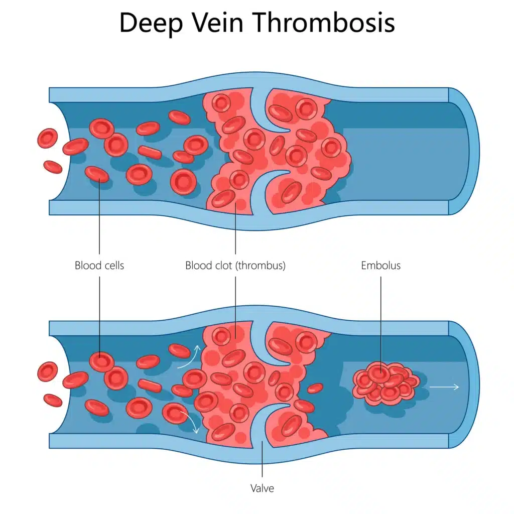
Deep Vein Thrombosis, commonly referred to as DVT, is a serious condition that occurs when a blood clot forms in one or more of the deep veins in your body, most often in the legs. This clot can restrict or completely block blood flow. The primary concern with DVT is the risk of a piece of the clot breaking off and traveling to the lungs, causing a potentially life-threatening pulmonary embolism.
The symptoms of DVT can include noticeable swelling in the affected leg, accompanied by pain, cramping, or soreness that typically begins in the calf area. The skin over the affected area may also appear red or discolored and feel warm to the touch. In some instances, DVT can occur without any obvious symptoms.
Diagnosis usually involves a physical examination and imaging tests, such as a Doppler ultrasound, to visualize the blood flow and locate any clots. Treatment for DVT primarily involves the use of blood-thinning medications to prevent the clot from growing and to reduce the risk of future clots. In more severe cases, minimally invasive procedures such as angioplasty may be used to open obstructed veins, and a filter may be placed in the vena cava to prevent clots from reaching the lungs. Research indicates that prompt treatment is essential for managing deep vein thrombosis and preventing long-term complications, like post-thrombotic syndrome.
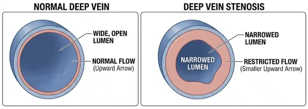
Deep Vein Stenosis is a condition characterized by the abnormal narrowing of the deep veins. This narrowing restricts the efficient flow of blood back to the heart, causing blood to pool in the extremities and increasing venous pressure. This condition can develop as a result of external compression from nearby anatomical structures or from scarring and damage caused by a previous DVT.
Patients with deep vein stenosis often experience chronic swelling, pain, and a feeling of heaviness or fatigue in the affected limb. Over time, the increased pressure can lead to skin changes and the development of open sores or ulcers.
To accurately diagnose deep vein stenosis, we utilize advanced imaging techniques such as ultrasound, CT scans, or MRIs. Once diagnosed, treatment often involves minimally invasive endovascular procedures. A common approach is angioplasty, where a small balloon catheter is inserted into the narrowed vein and inflated to widen it and restore blood flow. This is frequently followed by the placement of a venous stent, a small wire mesh tube that acts as a permanent scaffold inside the vein to maintain proper blood flow and prevent re-narrowing. According to research published in the Journal of Vascular Surgery: Venous and Lymphatic Disorders, venous stenting for iliac and vena caval occlusive disease is associated with high long-term patency rates and meaningful clinical improvement. These interventions are designed to alleviate symptoms and improve the patient’s quality of life.
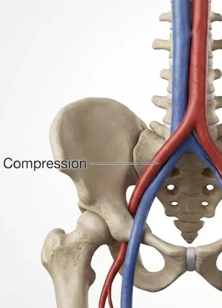
Iliac Vein Compression is a broader term that includes conditions like May-Thurner Syndrome, where the major veins in the pelvis are compressed, restricting blood flow from the lower extremities. This compression can be caused by overlying arteries, tumors, or other anatomical abnormalities. The resulting increased venous pressure can lead to significant discomfort and elevate the risk of blood clots.
The clinical presentation of iliac vein compression is similar to that of other venous outflow obstructions, including leg swelling, chronic pain, and a heavy sensation in the limbs. It can also contribute to the development of chronic venous insufficiency and, in some cases, pelvic congestion syndrome in women.
Managing iliac vein compression typically requires intervention to relieve the pressure on the vein. The primary treatment is angioplasty combined with venous stenting, a catheter-based procedure in which the compressed vein is first opened with a balloon and then held open with a wire mesh stent to restore and maintain adequate blood flow. Conservative measures such as compression stockings and leg elevation may be recommended for mild cases, but more significant compression generally requires this endovascular approach. Clinical data published in Endovascular Today has shown cumulative secondary patency rates of 93% at six years following iliac vein stenting, reflecting the durability of this treatment. If you are experiencing persistent leg swelling or pain, it is important to seek a professional evaluation. You can learn more about our diagnostic approach by reviewing our peripheral artery disease resources, as proper diagnosis is the first step toward effective treatment.
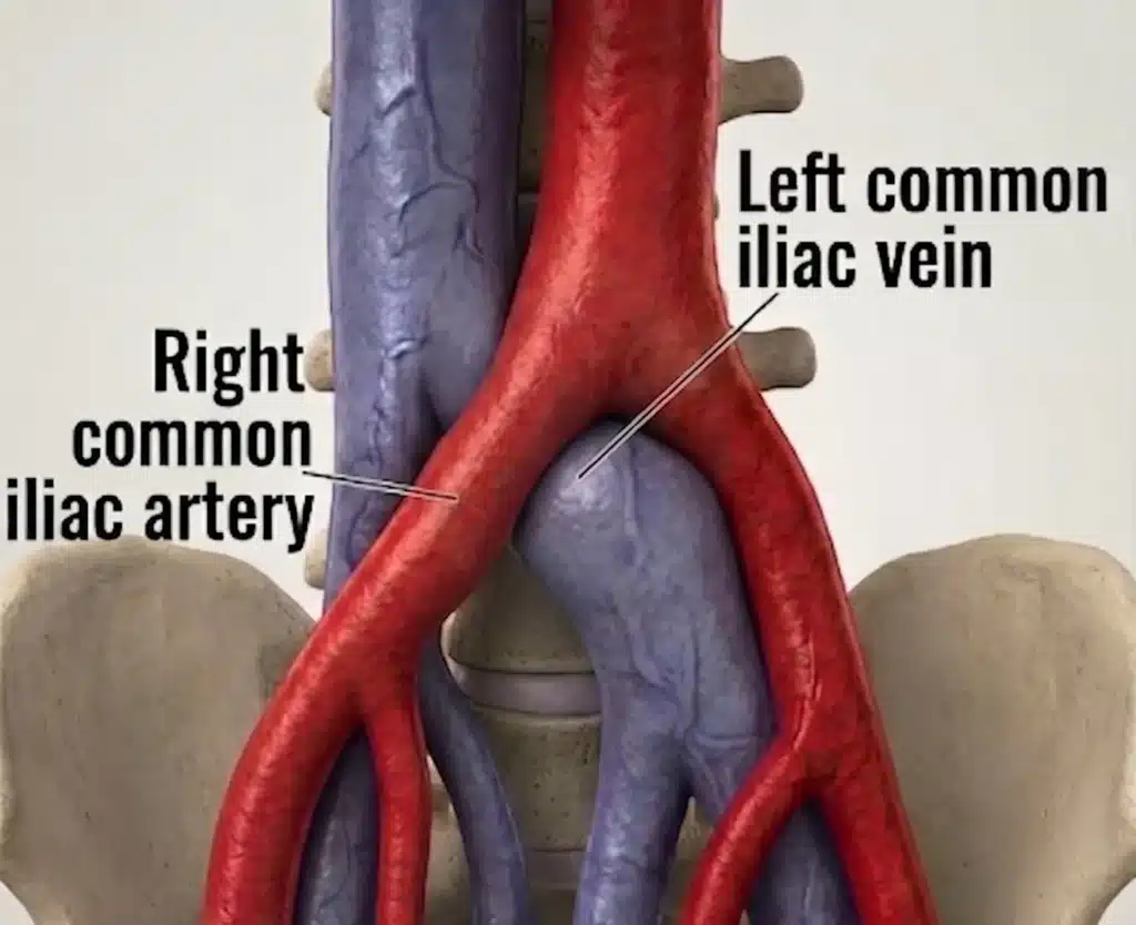
May-Thurner Syndrome, also known as iliac vein compression syndrome, is a specific anatomical condition where the right common iliac artery compresses the left common iliac vein against the spine in the pelvis. This compression hinders the return of blood from the left leg to the heart. While many people may have this anatomical variation without experiencing issues, it can significantly increase the risk of developing DVT in the left leg.
Many individuals with May-Thurner syndrome are asymptomatic until a complication arises. When symptoms do present, they typically affect the left leg and may include swelling, pain, heaviness, and the development of varicose veins.
Diagnosis involves a careful review of symptoms and imaging studies, including CT scans, MRIs, and intravascular ultrasound, to confirm the compression of the iliac vein. Treatment strategies focus on relieving the compression and restoring normal blood flow. For patients experiencing significant symptoms or those who have developed DVT, May-Thurner Syndrome is most commonly treated with angioplasty followed by iliac vein stenting.
During this procedure, a catheter is guided into the compressed vein, a balloon is inflated to open the narrowed segment, and a stent is placed to permanently hold the vein open. This approach is preferred over open surgery due to its shorter recovery time and strong clinical outcomes.
A venous ulcer, also known as a venous leg ulcer, is a chronic open wound that develops on the lower leg, most commonly near the ankle. These wounds are a direct result of long-standing venous insufficiency, where sustained high pressure in the veins damages the surrounding skin and tissue, preventing it from healing properly. Venous ulcers are among the most serious and debilitating complications of untreated venous disease, and they can be slow to heal without targeted treatment.
Venous ulcers typically present as shallow, irregularly shaped wounds with a red or yellow base, often surrounded by skin that appears discolored, hardened, or inflamed. The area around the ulcer may be swollen, itchy, or painful. Patients frequently report a history of varicose veins or chronic leg swelling prior to ulcer development, but many patients have no evidence of varicose veins. Because these wounds are driven by underlying venous disease, they tend to recur unless the root cause is addressed.
Diagnosing a venous ulcer involves a clinical assessment of the wound along with a duplex ultrasound to evaluate the venous system and confirm the presence of venous insufficiency or reflux. Treatment requires a two-part approach: managing the wound itself and correcting the underlying venous problem. Wound care may include specialized dressings, compression bandaging, and infection management. To address the underlying cause, procedures such as vein ablation or Varithena may be used to close the incompetent veins driving the increased venous pressure.
Research published in the Journal of Vascular Surgery: Venous and Lymphatic Disorders supports early endovenous intervention as a key factor in achieving ulcer healing and reducing the likelihood of recurrence. Treating the venous disease, not just the wound, is essential for lasting recovery.
If you are experiencing symptoms of a venous disease, such as chronic leg pain, swelling, varicose veins, or non-healing wounds, do not wait to seek professional care. Early diagnosis and treatment can prevent complications and significantly improve your quality of life. Our experienced team is here to provide the expert, compassionate care you deserve.